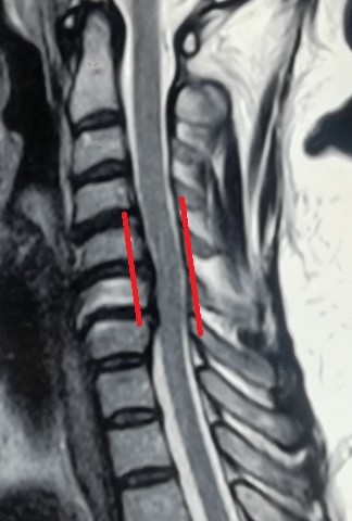
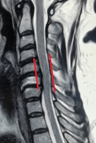
versatile herniated disc before surgery
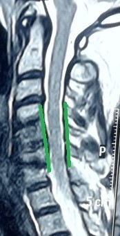
An image of an MRI scan of a 47-year-old patient suffering from partial quadriplegia due to multiple cervical disc herniation from the fourth cervical vertebra to the seventh. As shown in red, the location of the slip and the size of the effect resulting from pressure on the cervical spinal cord. And the image shaded in green after a surgical microscope surgery to remove the cartilage from the front and install alternative cervical cages after four months of surgery, as it shows the marrow liberation from pressure, which reflected on the patient's condition improvement.


