
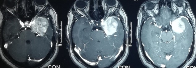
left temporal lobe meningioma
Brain magnetic resonance imaging, cross-sections of a 56-year-old woman suffering from visual impairment and severe headaches. As shown in the pictures in the upper row, the tumor is located in the left temporal lobe, pressing on the carotid artery and the optic nerve on the left side. And the bottom row is the x-ray view of the same patient four months after successful surgery to remove the tumor without any complications.

